Trending Topic

Musculoskeletal AI image
16 mins

Trending Topic

Developed by Touch
Mark CompleteCompleted
BookmarkBookmarked
Samantha Cooray, Alexander Deng, Tim Dong

There is much excitement about the deployment of artificial intelligence (AI) in healthcare, and the musculoskeletal field is no exception. In this article, we introduce some of the latest developments relating to osteoarthritis (OA), osteoporosis, rheumatoid arthritis (RA) (as an example of inflammatory arthritis), connective tissue disease (CTD), Ehlers–Danlos syndrome (EDS) and musculoskeletal surgical interventions. […]

Artificial Intelligence in Musculoskeletal Medicine: A Scoping Review

Samantha Cooray, Alexander Deng, Tim Dong, Salah AI Ghazal Hammouche, Matthew Quansah, Jordan Tsigarides, Nicholas Fuggle
Share
Facebook
X (formerly Twitter)
LinkedIn
Via Email
Mark CompleteCompleted
BookmarkBookmarked
Copy LinkLink Copied
Download as PDF
Published Online: Jul 5th 2024 touchREVIEWS in RMD. 2024;3(2):Online ahead of journal publication
Select a Section…
1

Abstract

Overview

In recent years, artificial intelligence (AI) has blossomed via the booming increase in computing power, access to large data sets and the development of novel data science techniques and architectures. This has led to a great interest in the clinical deployment of AI in healthcare. In this review, we look particularly at the field of musculoskeletal medicine and have curated the latest progress in AI and machine learning in the areas of inflammatory arthritis, vasculitis and connective tissue diseases, osteoporosis, Ehlers–Danlos syndrome, osteoarthritis and musculoskeletal surgical interventions. We also highlight the challenges and pitfalls as clinical AI moves forward.

Keywords
2

Article

There is much excitement about the deployment of artificial intelligence (AI) in healthcare, and the musculoskeletal field is no exception. In this article, we introduce some of the latest developments relating to osteoarthritis (OA), osteoporosis, rheumatoid arthritis (RA) (as an example of inflammatory arthritis), connective tissue disease (CTD), Ehlers–Danlos syndrome (EDS) and musculoskeletal surgical interventions.

Artificial intelligence

AI is the ability of machines to imitate human behaviour. Machine learning (ML) is a branch of AI that uses computer algorithms to automatically learn from input data and through the identification of patterns to make accurate predictions with minimal human intervention. ML algorithms do not rely on a predetermined equation as a model but adaptively improve their performance as more data are provided.

ML is increasingly being used in medical applications to independently and efficiently carry out specified tasks, such as image processing in radiology and histopathology and the identification of disease markers, and in the field of drug discovery.

The process by which ML algorithms learn is called ‘training’, and the training methods are classified as supervised, unsupervised or reinforcement learning. Supervised learning involves training algorithms to predict future values through learning patterns from labelled input data, for example, accurately predicting the opacification on a chest X-ray through training with images that are labelled as normal or abnormal.

Supervised learning algorithms use classification, regression and forecasting approaches. In classification models, the algorithm must draw a conclusion from the observed values and determine to which category new observations belong. In regression tasks, the ML algorithm must estimate and understand the relationships among variables. Forecasting is the process of making predictions about the future based on past and present data and is commonly used to analyse trends.

In unsupervised learning, the input data are not assigned or labelled. Instead, the algorithm infers the underlying patterns and relationships within the input data, which is useful, for example, in finding patterns in blood biomarkers that are associated with a particular disease state. Clustering is a type of unsupervised learning that involves grouping sets of similar data based on the defined criteria. It is useful for segmenting the data into groups and performing analysis on each data set to find patterns. Dimension reduction is another type of unsupervised learning that gradually reduces the number of variables being considered to find the exact information required. In reinforcement learning, the algorithm achieves a specified goal by interacting with an environment through trial and error. This type of method is commonly applied in the field of robotic surgery.

Examples of ML algorithms are shown in Figure 1 and described in greater depth in the glossary (online supplementary file 1).

Figure 1: Schematic representations of machine learning and deep learning methods

Figure 1: Schematic representations of machine learning and deep learning methods

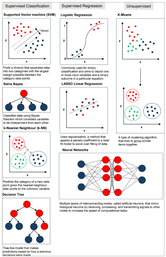
LASSO = least absolute shrinkage and selection operator.

Inflammatory arthritis

Inflammatory arthritides are characterized by joint inflammation, leading to clinical signs, such as pain, swelling, heat and erythema. This collection of rheumatological conditions encompasses those causing inflammation due to various aetiologies, including autoimmunity, crystals and reactive processes. In this review, the focus will be on RA as a vital example of inflammatory arthritis.

RA is a chronic, systemic autoimmune disease leading to inflammatory arthritis, typically involving the small joints that can result in joint destruction and severe functional impairment.1 RA is a heterogeneous disease with a wide variation in the age of onset, degree of joint involvement and severity. The diagnosis is based on clinical criteria, and there is no definitive diagnostic test available to differentiate RA from other inflammatory joint diseases.2 The aim of treatment for RA is to reduce inflammation and prevent joint destruction, and it includes corticosteroids, conventional disease-modifying anti-rheumatic drugs (DMARDs) and an ever-expanding list of targeted biological therapies.3 Patients have variable response and remission rates to different treatment strategies, and identifying responders from non-responders is difficult. However, early diagnosis and prompt initiation of treatment are important in the prevention of inflammatory joint destruction. AI and ML are increasingly being used to address the unmet needs in the diagnosis, monitoring and treatment of RA, and in this section, we review the latest developments.4

Machine learning in inflammatory arthritis diagnosis

AI and ML for diagnostic purposes use the data from electronic health records (EHRs), diagnostic biomarkers and imaging modalities to identify patients with RA at an early stage of the disease.

Electronic health records

The phenotypic identification of RA using data from EHRs was first carried out in 2010 using a support vector machine (SVM) model, which accurately predicted RA disease activity from the data containing specific terms (area under the curve [AUC] >0.90).5 The comparison of different ML methods using a 10-fold cross-validation from thousands of EHRs showed that an SVM model performed best in identifying patients with RA in <7 s (an AUC of 0.98 and a positive predictive value [PPV] of 0.94).6 The model compared favourably with manual methods.7 A random forest method and a decision tree model identified the best predictors of RA, such as local coding for RA; clinical features, such as nodules, seropositivity and arthropathy and drugs, such as prednisolone, methotrexate, sulfasalazine and leflunomide, from EHRs, with a PPV of 85.6%, specificity of 94.6% and sensitivity of 86.2%.8

Molecular markers

Random forest, logistic regression and other statistical models have differentiated patients with RA from controls using gene expression signatures, with an AUC of 0.96.9,10 Artificial neural networks (ANNs) have successfully distinguished RA from OA and psoriatic arthritis (PsA, another form of inflammatory arthritis) using serum protein profiles identified through mass spectrometry and immunoassays, although the AUC varies significantly.11–13

Imaging techniques and histopathology

Supervised and unsupervised algorithms have been used, together with imaging techniques, for RA diagnosis. ANN models trained and tested on hand radiographs accurately classified RA with an AUC of 0.97, a sensitivity of 90.7% and a specificity of 92.6%.14,15 Larger data sets have allowed the effective differentiation of RA and OA.16 ANNs also identified synovial proliferation in joint ultrasound with an AUC of 0.863–0.886, depending on the joint area sampled.17,18 A systemic review and meta-analysis of computer-assisted magnetic resonance imaging (MRI) showed a good correlation with manual methods in RA diagnosis.19 Thermal imaging of diurnal joint temperatures has been studied, with quantum SVM models used to improve the accuracy.20,21 Scoring algorithms based on features, such as inflammatory infiltrates and hyperplasia in stained synovial tissue, have demonstrated considerable efficacy in differentiating RA, OA and PsA from normal tissue (AUC 0.87–0.98).22,23

Disease progression and disease activity

Once RA is diagnosed, an ongoing assessment is vital to monitor the disease activity. An ordinal regression-based ML model developed to estimate the Clinical Disease Activity Index (CDAI) scores was trained and validated on 11,985 patient notes with CDAI scores from the OM1 RA registry (OM1® real-world data and AI for research [OM1, Boston, MA, USA).24 The model had a PPV of 0.80, a negative predictive value of 0.84 and an AUC of 0.88 when evaluating the performance using the binarized (negative CDAI score ≤10 or positive CDAI score ≥10.1) version of the outcome.

Predicting treatment response

ML techniques have shown promise in the evaluation of treatment response to DMARDs and biologics. Least absolute shrinkage and selection operator and random forest methods showed that patients with RA with a low baseline Disease Activity Score in 28 joints (DAS28)-erythrocyte sedimentation rate (ESR) score, positive anti-citrullinated protein antibody and the Health Assessment Questionnaire ≤2 were most likely to respond well to methotrexate (AUC of 0.68 and 0.79, respectively).25 Conversely, a DAS28-ESR score (>3.2) at 3 months post-methotrexate treatment predicted a poor therapeutic response with different methods.26 Elastic net models demonstrated a 3.6-fold better response to anti-tumour necrosis factor (TNF) biologics in males, and using genetic data, carriers of the resistin gene rs3219177 polymorphism had a sixfold better response than non-carriers. In a subsequent study, carriers of the TLR9 gene rs352139 polymorphism were found to have a fivefold better response to anti-TNF than non-carriers.27,28 ML using biomarker signatures in blood or synovial tissue has reliably predicted responses to other biologics (e.g. rituximab, tocilizumab and sarilumab).29–31

Vasculitis and connective tissue disease

Vasculitis is a complex group of rare rheumatic diseases characterized by inflammation of blood vessels that present considerable diagnostic and therapeutic challenges. Recent studies using AI techniques have attempted to support earlier diagnosis, treatment optimization and prognosis prediction.32–34

The diagnosis of vasculitis relies upon the recognition of characteristic clinical features often alongside the use of imaging or biopsy. ML techniques show great promise for supporting this process, with studies using natural language processing to differentiate vasculitis from clinical notes, deep learning algorithms (such as U-Net) to detect features on medical imaging and supervised learning approaches to identify predictive biomarkers detectable from blood samples.35–41

Treatment typically includes induction through high-dose corticosteroids followed by maintenance with ‘steroid-sparing’ agents. However, variability in treatment response combined with common side effects makes management fraught with difficulty. ML has been used to address treatment challenges through supervised algorithms (including random forest and light gradient boosting models) to predict relapse after glucocorticoid tapering in giant cell arteritis and the development of models predicting resistance to intravenous immunoglobulin therapy in Kawasaki’s disease.32,42,43

The risk of morbidity following a diagnosis of vasculitis is significant, and understanding the prognostic risk factors is essential to tailor management strategies. Emerging ML-based tools show promise, especially in predicting eye diseases from clinical data (using ensemble algorithms, such as XGBoost) or angiography imaging and risk of renal damage in immunoglobulin A (IgA) vasculitis based on patients’ clinical features.44–46

With only 26 primary research studies published over the last 20 years using AI methods in vasculitis, and the majority investigating Kawasaki’s disease (n=12), there is still a dearth of available evidence despite many studies showing highly promising results.47–57

Autoimmune CTDs are systemic in nature and characterized by a variety of phenotypes. ML techniques have begun to revolutionize the study of these varied diseases, providing tools for improving diagnostic approaches, the classification of disease subtypes and predicting factors associated with treatment response and the development of disease-related complications.

CTDs present a diagnostic challenge for clinicians, and many studies using ML focus on demonstrating accurate tools to support the diagnostic process. There are encouraging studies investigating gene expression data as novel biomarkers in CTDs, but perhaps, the approaches closer to clinical implementation are those using deep learning to support and automate the interpretation of clinical data (including point-of-care tests, radiological imaging and biopsies).58–71 An interesting example includes the use of deep learning to differentiate systemic sclerosis from primary Raynaud’s syndrome using a low-cost digital nailfold capillaroscope, an underused tool in which AI may support more widespread use.63

The spectrum of CTD presentations highlights the importance of classification, subtype definition and tailored treatment strategies. Data mining and supervised approaches have been used to discriminate subtypes of Sjögren’s syndrome and lupus based on clinical data, in addition to multiple studies investigating the classification of myositis using electromyography and MRI data.72–76

Many of the AI-based studies on CTDs aim to predict outcomes and treatment responses to inform a personalized management approach. Multiple ML approaches have been used to predict complications and comorbidities in lupus (including nephritis), pulmonary involvement in systemic sclerosis and response to immunomodulatory treatments.77–97

Osteoporosis

Osteoporosis is characterized by reduced bone mineral density (BMD), leading to a predisposition to a fragility fracture. The diagnosis is complicated by the absence of symptoms or clinical signs and is reliant on measures of BMD. BMD is primarily derived from dual-energy X-ray absorptiometry (DXA) as a single parameter, and it is this measurement that informs the densitometric diagnosis of osteoporosis, defined as a BMD T-score of ≤-2.5. AI techniques have been used to interrogate ‘omic’ data sets to identify biomarkers and therapeutic targets in osteoporosis, as they have in most major disease areas; however, the focus of this review will be on the deployment of AI on skeletal imaging (computer vision) in the field of osteoporosis.98,99

The major aims of computer vision in osteoporosis are as follows:

  • use DXA images to improve fracture prediction beyond BMD

  • opportunistically measure BMD from routinely performed imaging

  • opportunistically identify vertebral fractures from routinely performed imaging

  • derive bone microarchitecture features.

It is worth noting that, in the field of osteoporosis, the outcome of interest can be either the diagnosis of osteoporosis or the assessment of fracture risk. Although the former was vital for defining the disease and developing the field, the measures of the latter (fracture risk) are used to adjudicate treatment thresholds via fracture risk-prediction tools, including The Fracture Risk Calculator (FRAX®), which provides a 10-year estimation of major osteoporotic risk and hip fracture risk.100 This is, in part at least, as over half of fragility fractures occur at a normal BMD and so a pure focus on the diagnosis of osteoporosis would miss a large proportion of those at risk of fracture. Indeed, fractures are associated with high morbidity and mortality, surpassing simply the densitometric diagnosis of osteoporosis.

As mentioned earlier, the DXA scan is performed to provide a measure of BMD (via levels of beam attenuation), but the rest of the DXA image (of the hips and anterior–posterior spine) is neglected unless a lateral image of the spine is acquired as a vertebral fracture assessment. These acquired, but underused, DXA images are ripe for computer vision analyses.

Indeed, a meta-analysis of seven studies that attempt to improve the diagnosis of osteoporosis via hip DXA images found a pooled sensitivity of 0.844 (95% confidence interval [CI], 0.79–0.89), a pooled specificity of 0.781 (95% CI, 0.73–0.82) and a summary of receiver operating characteristic AUC of 0.878.101 Although these results are encouraging, DXA imaging is limited by quality due to artefacts of soft tissue and degenerative sclerosis and, in a clinical setting, is only performed to assess BMD. Plain radiographs and computed tomography (CT) are imaging modalities that are used for a plethora of clinical indications and often contain skeletal elements. These images could all contribute to the assessment of skeletal health opportunistically.

This has led to studies showing that abdominal CT can predict lumbar spine BMD (AUC 0.96–0.97), chest CT scans (performed for lung cancer screening) can discriminate osteoporosis from normal participants (AUC 0.97) and lateral spine X-rays can identify osteoporosis, though with a lower degree of accuracy (AUC 0.85).102,103

Vertebral fractures are often missed on routine imaging, and hence, models have been developed using deep learning (amidst other methods) to identify vertebral fractures from lateral spine radiographs (AUC 0.93) and sagittal reconstructions of CT images.104,105

Bone is composed of two compartments, cortical and trabecular, and information regarding these compartments can provide more detailed ‘bone microarchitectural’ measures of bone strength and resistance to fracture than simply BMD alone. This microarchitecture can be derived from specialist scans currently largely used in research settings, including high-resolution peripheral quantitative CT (HR-pQCT). Computer vision analyses of HR-pQCT have demonstrated excellent discrimination for prevalent fractures with an AUC of 0.85–0.97 using a gradient boosting machine and statistical shape mapping.106–108 What is more, there are efforts, using AI, to derive some microarchitectural features (though only from the trabecular compartment) from the DXA imaging.109

In conclusion, computer vision analyses have improved the identification of fracture risk on DXA images, developed the opportunistic screening of BMD and vertebral fractures and shown promise for deriving bone microarchitectural properties from clinically applicable imaging modalities.

Ehlers–Danlos syndrome

EDSs are a group of inherited disorders of connective tissue. Within this group, there are 14 recognized EDS subtypes, 13 of which are genetically characterized. The clinical presentation commonly includes features such as joint hypermobility, skin hyperextensibility, abnormal wound healing and widespread pain. Similar to connective tissue disorders, EDSs are not solely disorders of the musculoskeletal system but are complex multi-system disorders due to the role of connective tissue throughout the body. AI tools in development may eventually assist in the diagnosis of EDS, with two current examples being video assessment of joint hypermobility and the classification of rare diseases via analysis of patient-created pain drawings.

A thematic clinical feature of EDS is generalized joint hypermobility. The clinical finding of joint hypermobility is a core diagnostic feature measured through the Beighton examination completed using a goniometer. However, the use of a goniometer is uncommon in actual clinical practice, and evidence of inter-rater reliability in the Beighton score reporting is limited or conflicting.110 Mittal et al. have devised a method to apply computer vision to smartphone camera footage of patients, with the goal of creating a more objective, validated and scalable method for assessing hypermobility.111 This provides an excellent case study to highlight the design of research in the field of musculoskeletal AI.

The footage will be analysed using ML algorithms from the existing open-source human pose-estimation models, which use the video input to create an output of the location and angles of joints in the body. Pose-estimation models lack training data on generalized joint hypermobility, potentially creating out-of-distribution errors for the pose libraries. The authors plan to address this by additional fine-tuning of the models with manually annotated examples of hyperextended joints. The method is planned to be validated compared with the ground truth of expert clinicians using the goniometer-validated assessment.

The outcomes of the pre-registered trial are awaited in 2024 (Assessing the Feasibility of a Smartphone-based, Machine Learning Visual Imaging Application for Assessment of Hyperextensibility of Peripheral Joints in Ehlers Danlos Syndrome; ClinicalTrials.gov identifier: NCT05366114), and the authors anticipate possible use cases as a triage tool for specialized EDS clinic referrals.112

Chronic pain is another thematic clinical feature of EDS. There are many aetiologies of chronic pain, including other rare diseases, and management is informed by the underlying cause. Emmert et al. developed an algorithm trained to distinguish between patterns of chronic pain caused by different rare diseases.113 The input data to the algorithm were pain drawings. Pain drawings are made by patients who mark where they experience pain on a 2D outline of a human body to create a visual representation of their pain (instead of relying on language). The authors used an open-source platform ‘Pain2D’ to collect the data from patients with specific known rare disease diagnoses involving chronic pain: EDS, GuillainBarré syndrome, facioscapulohumeral muscular dystrophy, proximal myotonic myopathy and a control group with non-specific chronic pain. An overall pain profile for each disease category was created using this platform by calculating the frequency with which each pixel in the pain drawing was marked over all instances of each diagnostic category. Pain profiles summarizing different diagnostic categories were then compared with the known cases (leave-one-out cross-validation) by a mathematical coefficient of similarity – the highest-scoring pain profile was chosen as the output classification. This method classified EDS among the five diagnostic categories with a sensitivity of 64.4% and a specificity of 88.7%. Significant limitations include the absence of pain-drawing data from other diagnostic categories of chronic pain and small data sets within the existing categories that precluded the use of deep learning by convolutional neural networks (CNNs).

It is anticipated that these study areas will be developed and built upon but provide good examples of AI study designs that have the potential to be used across musculoskeletal disorders.

Osteoarthritis

OA is a common joint disease affecting millions of people throughout the world but is plagued by a disconnect between clinical and radiographic diseases. In recent years, ML and AI techniques have shown promise for enhancing OA diagnosis, care and management.114 This section seeks to give readers an overview of the most recent developments in ML and AI applications in the field of OA.

Early diagnosis and disease progression detection

Early diagnosis and detection algorithms use a variety of data sources, including patient-reported outcomes and medical imaging (X-rays and MRI scans), to detect the patterns and signals linked to the disease. This can take different approaches, but one study combined clinical, imaging and demographic features using a multi-modal feature integration method using an L-1 normalization approach to minimize the number of irrelevant features from each modality.115 Early OA symptoms can be recognized so that therapies can start sooner, possibly slowing the disease’s progression.

To develop predictive models for OA progression, ML algorithms have been trained on big data sets combining patient demographics, clinical data and imaging results. These models determine the chance that an illness will progress and assist physicians in selecting the best course of action, potentially improving patient outcomes via a clinical decision support system. One research presented a CNN model referred to as the YOLOv3 tiny-based object identification approach to automatically detect and categorize knee OA according to the Kellgren–Lawrence (KL) classification scheme.116 The strategy used two models, one for classifying severity (progression) and the other for distinguishing between healthy and osteoarthritic knees. The CNN model sought to assist radiologists and orthopaedic surgeons by correctly identifying and classifying knee OA across various stages. Another research introduced DenseNet169 as a model for the effective diagnosis of knee OA.117 It compared DenseNet169’s performance with five other deep learning systems and highlighted its efficacy in finding knee OA and determining its severity through multi-classification (accuracy of 95%, sensitivity of 88.77%, specificity of 95.41% and precision of 87.08%) and binary classifications (accuracy of 93.78%, sensitivity of 91.29%, specificity of 87.57% and accuracy of 89.27%). The suggested methodology showed potential in relieving radiologists’ burden, enabling early diagnosis and saving time and expense related to knee OA diagnosis. It used a weighted training approach and incorporated gradient-weighted class activation mapping, a heatmap-based algorithm that deep learning models used to visualize and decipher the significant areas of an image (using gradients in the final convolutional layer) that influence the model’s prediction.

Treatment plans for people with OA have been improved using ML and AI techniques. Algorithms can recommend personalized treatment programmes that are suited to each patient’s needs by examining patient data, including medical history, genetic factors and lifestyle variables. By identifying the most efficient interventions and minimizing the process of trial and error while choosing treatments, this strategy seeks to improve patient outcomes.

One study found that AI-aided clinician diagnostic ratings have a stronger correlation with the total KL scores and Knee Injury and Osteoarthritis Outcome Scores than those by unaided physicians, increasing the consistency and precision of OA diagnosis.118 The KL score appears to be a limited tool on its own, with only a modest relationship between clinical severity and radiographic assessments. AI assistance has the potential to address the disconnect between radiological OA severity and clinical symptoms, resulting in more precise detection and better patient care.

The use of wearable and sensor technologies in conjunction with ML algorithms enables remote monitoring of patients with OA. These devices can monitor joint motion, levels of activity and other important factors. Clinicians can provide real-time feedback and useful insights into patient situations by analysing the acquired data, enabling proactive management and intervention. For monitoring human movement, wearable sensors provide an affordable and practical alternative to optical motion capture.119 Large amounts of real-world data can be provided by them, enabling remote monitoring, resolving time restrictions in the delivery of remote treatment and encouraging patient interaction. For movement analysis, accelerometers, gyroscopes and magnetometers are frequently combined into inertial measurement units for cost-efficiency and usability in large cohorts. For people with hip or knee OA, including those who have had joint replacement surgery, wearable sensors hold great promise for improving research and care.

Another study validated a remote patient monitoring system for 25 patients with OA undergoing total knee arthroplasty using a wearable knee sleeve and smartphone applications.120 The sleeve accurately measured mobility, range of motion, patient-reported outcome measures, opioid compliance and home exercise compliance. Such studies demonstrate the potential of AI-augmented remote patient monitoring for evaluating hip and knee arthroplasty recovery and rehabilitation.

In OA management, persistent challenges remain around the quality and accessibility of data. Obtaining comprehensive data sets spanning diverse patient profiles, multiple data modalities and longitudinal records remains a hurdle, essential for training reliable ML models. Another critical challenge lies in the interpretability and explainability of these models; understanding the rationale behind their diagnoses or treatment suggestions is crucial for building trust among healthcare professionals and patients. Additionally, ensuring the ethical use of sensitive medical information while maintaining patient privacy poses an ongoing concern. Moreover, integrating these advanced technologies safely into clinical workflows and healthcare systems presents a significant challenge for wider adoption and practical implementation in real-world settings.

Musculoskeletal surgical interventions

The sphere of surgical intervention has notably benefitted from the introduction of AI-aided technologies, which present the potential to amplify several aspects of surgical care.121–128 The purview of these tools encompasses diagnosis and planning to intraoperative guidance.121–128 A salient advantage rests in their capacity to aid clinicians in the early detection of musculoskeletal disorders, such as OA and soft tissue pathologies.123,127,129,130 Furthermore, the competency of AI in the precise identification of fractures and distinguishing between diverse types of spondylitis parallels that of experienced radiologists potentially enabling faster management.131–133

In the planning phase, AI resources are able to deliver patient-specific preoperative plans, thereby minimizing the necessity for surgeon-delivered modifications to the surgical plan.121,123,134 These resources can also construct 3D models of patient anatomy, providing orthopaedic surgeons with a holistic and detailed visual representation of the surgical area.124,125 In the operating theatre, AI’s role expands to offering real-time feedback and assistance in navigating complex anatomical structures.124,126,135,136

Postoperative care has similarly been affected by the advent of AI, aiding in the identification of implant positioning and type, forecasting potential implant failure and assisting in planning for future surgical interventions.122,137,138 AI-assisted tools have manifested as an efficacious solution to the common difficulties in postoperative CT and MRI associated with metal-related image artefact and distortion.139 AI can also help in determining follow-up recommendation plans based on analysing the radiological report.140

Challenges

The field of musculoskeletal AI is filled with optimism, but there are substantial challenges that must be addressed to enable safe, unbiased and equitable deployment in clinical practice.141–143

Most current ML methods use supervised methods using large data sets, which need manual checks and large amounts of data. This means that the accuracy of the models relies on the quality of the input data. Quality control measures are, therefore, required, and these need time-consuming human input.

The integration of AI algorithms into the clinical workflow must be performed carefully and thoughtfully and be congruent with the existing information technology infrastructure.141–143 The replication of AI studies on ‘real-life’ data remains problematic due to the sporadic availability of training data and code and must be addressed moving forward and is sometimes neglected in favour of model performance.142

The data for future AI research must be suitably anonymized with consideration of patient privacy and ethical considerations.142 The patients’ preference for human experts’ diagnoses accentuates the importance of cultivating appropriate levels of trust in AI applications. Additionally, considerable challenges persist in data availability and model validation, especially for rare musculoskeletal disorders and diseases (including vasculitis and CTD). The regulation in AI is developing and is vital to ensure that these tools are created, maintained and used appropriately in clinical practice.142,144,145

Conclusions

AI within the musculoskeletal field harbours immense benefits, and as detailed earlier, a great deal of work has been performed and is ongoing. As research and development progress, AI has the potential to become an increasingly vital component of musculoskeletal care. However, the above challenges must be considered of utmost importance as the field moves forward.146 While AI embodies the promise of enhanced patient care, cognizance of the related challenges and ethical considerations is indispensable for wider acceptance and implementation of AI in the musculoskeletal field.

3

References

List View
Grid View
1
Copy DOIDOI Copied
Visit DOI Link

 Momtazmanesh SNowroozi ARezaei NArtificial intelligence in rheumatoid arthritis: Current status and future perspectives: A state-of-the-art reviewRheumatol Ther2022;9:1249304DOI10.1007/s40744-022-00475-4.

2
Copy DOIDOI Copied
Visit DOI Link

 Gravallese EMFirestein GSRheumatoid arthritis – Common origins, divergent mechanismsN Engl J Med. 2023;388:52942. DOI10.1056/NEJMra2103726.

3
Copy DOIDOI Copied
Visit DOI Link

 Prasad PVerma SSurbhiet alRheumatoid arthritis: Advances in treatment strategiesMol Cell Biochem. 2023;478:6988. DOI10.1007/s11010-022-04492-3.

4
Copy DOIDOI Copied
Visit DOI Link

 Kedra JDavergne TBraithwaite Bet alMachine learning approaches to improve disease management of patients with rheumatoid arthritis: Review and future directionsExpert Rev Clin Immunol2021;17:131121. DOI10.1080/1744666X.2022.2017773.

5
Copy DOIDOI Copied
Visit DOI Link

 Carroll RJEyler AEDenny JCNaive electronic health record phenotype identification for rheumatoid arthritisAMIA Annu Symp Proc2011;2011:18996.

6
Copy DOIDOI Copied
Visit DOI Link

 Maarseveen TDMeinderink TReinders MJTet alMachine learning electronic health record identification of patients with rheumatoid arthritis: Algorithm pipeline development and validation studyJMIR Med Inform. 2020;8:e23930. DOI10.2196/23930.

7
Copy DOIDOI Copied
Visit DOI Link

 Maarseveen TDMaurits MPNiemantsverdriet Eet alHandwork vs machine: A comparison of rheumatoid arthritis patient populations as identified from EHR free-text by diagnosis extraction through machine-learning or traditional criteria-based chart reviewArthritis Res Ther2021;23:174DOI10.1186/s13075-021-02553-4.

8
Copy DOIDOI Copied
Visit DOI Link

 Zhou S-MFernandez-Gutierrez FKennedy Jet alDefining disease phenotypes in primary care electronic health records by a machine learning approach: A case study in identifying rheumatoid arthritisPLoS One. 2016;11:e0154515. DOI10.1371/journal.pone.0154515.

9
Copy DOIDOI Copied
Visit DOI Link

 Liu JChen NA 9 mRNAs-based diagnostic signature for rheumatoid arthritis by integrating bioinformatic analysis and machine-learningJ Orthop Surg Res2021;16:44DOI10.1186/s13018-020-02180-w.

10
Copy DOIDOI Copied
Visit DOI Link

 Long NPPark SAnh NHet alEfficacy of integrating a novel 16-gene biomarker panel and intelligence classifiers for differential diagnosis of rheumatoid arthritis and osteoarthritisJ Clin Med2019;8:50DOI10.3390/jcm8010050.

11
Copy DOIDOI Copied
Visit DOI Link

 Chocholova EBertok TJane Eet alGlycomics meets artificial intelligence – Potential of glycan analysis for identification of seropositive and seronegative rheumatoid arthritis patients revealedClin Chim Acta2018;481:4955DOI10.1016/j.cca.2018.02.031.

12
Copy DOIDOI Copied
Visit DOI Link

 Heard BJRosvold JMFritzler MJet alA computational method to differentiate normal individuals, osteoarthritis and rheumatoid arthritis patients using serum biomarkersJ R Soc Interface. 2014;11:20140428. DOI10.1098/rsif.2014.0428.

13
Copy DOIDOI Copied
Visit DOI Link

 Mc Ardle AKwasnik ASzentpetery Aet alIdentification and evaluation of serum protein biomarkers that differentiate psoriatic arthritis from rheumatoid arthritisArthritis Rheumatol2022;74:8191DOI10.1002/art.41899.

14
Copy DOIDOI Copied
Visit DOI Link

 Mate GSKureshi AKSingh BKAn efficient CNN for hand X-ray classification of rheumatoid arthritisJ Healthc Eng2021;2021:6712785DOI10.1155/2021/6712785.

15
Copy DOIDOI Copied
Visit DOI Link

 Üreten KErbay HMaraş HHDetection of rheumatoid arthritis from hand radiographs using a convolutional neural networkClin Rheumatol2020;39:96974DOI10.1007/s10067-019-04487-4.

16
Copy DOIDOI Copied
Visit DOI Link

 Üreten KMaraş HHAutomated classification of rheumatoid arthritis, osteoarthritis, and normal hand radiographs with deep learning methodsJ Digit Imaging2022;35:1939DOI10.1007/s10278-021-00564-w.

17
Copy DOIDOI Copied
Visit DOI Link

 Andersen JKHPedersen JSLaursen MSet alNeural networks for automatic scoring of arthritis disease activity on ultrasound imagesRMD Open2019;5:e000891DOI10.1136/rmdopen-2018-000891.

18
Copy DOIDOI Copied
Visit DOI Link

 Wu MWu HWu Let alA deep learning classification of metacarpophalangeal joints synovial proliferation in rheumatoid arthritis by ultrasound imagesJ Clin Ultrasound2022;50:296301DOI10.1002/jcu.23143.

19
Copy DOIDOI Copied
Visit DOI Link

 Haj-Mirzaian AKubassova OBoesen Met alComputer-assisted image analysis in assessment of peripheral joint MRI in inflammatory arthritis: A systematic review and meta-analysisACR Open Rheumatol2022;4:72134. DOI10.1002/acr2.11450.

20
Copy DOIDOI Copied
Visit DOI Link

 Ahalya RKSnekhalatha UDhanraj VAutomated segmentation and classification of hand thermal images in rheumatoid arthritis using machine learning algorithms: A comparison with quantum machine learning techniqueJ Therm Biol2023;111:103404DOI10.1016/j.jtherbio.2022.103404.

21
Copy DOIDOI Copied
Visit DOI Link

 Bardhan SBhowmik MK2-stage classification of knee joint thermograms for rheumatoid arthritis prediction in subclinical inflammationAustralas Phys Eng Sci Med2019;42:25977DOI10.1007/s13246-019-00726-9.

22
Copy DOIDOI Copied
Visit DOI Link

 Slansky ELi JHäupl Tet alQuantitative determination of the diagnostic accuracy of the synovitis score and its componentsHistopathology2010;57:43643DOI10.1111/j.1365-2559.2010.03641.x.

23
Copy DOIDOI Copied
Visit DOI Link

 Mehta BGoodman SDiCarlo Eet alMachine learning identification of thresholds to discriminate osteoarthritis and rheumatoid arthritis synovial inflammationArthritis Res Ther2023;25:31DOI10.1186/s13075-023-03008-8.

24
Copy DOIDOI Copied
Visit DOI Link

 Spencer AKBandaria JLeavy MBet alValidation of a machine learning approach to estimate Clinical Disease Activity Index scores for rheumatoid arthritisRMD Open2021;7:e001781DOI10.1136/rmdopen-2021-001781.

25
Copy DOIDOI Copied
Visit DOI Link

 Duong SQCrowson CSAthreya Aet alClinical predictors of response to methotrexate in patients with rheumatoid arthritis: A machine learning approach using clinical trial dataArthritis Res Ther. 2022;24:162. DOI10.1186/s13075-022-02851-5.

26
Copy DOIDOI Copied
Visit DOI Link

 Gosselt HRVerhoeven MMABulatović-Ćalasan Met alComplex machine-learning algorithms and multivariable logistic regression on par in the prediction of insufficient clinical response to methotrexate in rheumatoid arthritisJ Pers Med2021;11:44DOI10.3390/jpm11010044.

27
Copy DOIDOI Copied
Visit DOI Link

 Kim WJin Oh SThi Trinh Net alEffects of RETN polymorphisms on treatment response in rheumatoid arthritis patients receiving TNF-alpha inhibitors and utilization of machine-learning algorithmsInt Immunopharmacol. 2022;111:109094. DOI10.1016/j.intimp.2022.109094.

28
Copy DOIDOI Copied
Visit DOI Link

 Kim WKim THOh SJet alAssociation of TLR 9 gene polymorphisms with remission in patients with rheumatoid arthritis receiving TNF-alpha inhibitors and development of machine learning modelsSci Rep. 2021;11:20169. DOI10.1038/s41598-021-99625-x.

29
Copy DOIDOI Copied
Visit DOI Link

 Johansson FDCollins JEYau Vet alPredicting response to tocilizumab monotherapy in rheumatoid arthritis: A real-world data analysis using machine learningJ Rheumatol2021;48:136470DOI10.3899/jrheum.201626.

30
Copy DOIDOI Copied
Visit DOI Link

 Rehberg MGiegerich CPraestgaard Aet alIdentification of a rule to predict response to sarilumab in patients with rheumatoid arthritis using machine learning and clinical trial dataRheumatol Ther2021;8:166175. DOI10.1007/s40744-021-00361-5.

31
Copy DOIDOI Copied
Visit DOI Link

 Rivellese FSurace AEAGoldmann Ket alRituximab versus tocilizumab in rheumatoid arthritis: Synovial biopsy-based biomarker analysis of the phase 4 R4Ra randomized trialNat Med2022;28:125668DOI10.1038/s41591-022-01789-0.

32
Copy DOIDOI Copied
Visit DOI Link

 Sunaga YWatanabe AKatsumata Net alA simple scoring model based on machine learning predicts intravenous immunoglobulin resistance in Kawasaki diseaseClin Rheumatol2023;42:135161DOI10.1007/s10067-023-06502-1.

33
Copy DOIDOI Copied
Visit DOI Link

 Duff LMScarsbrook AFRavikumar Net alAn automated method for artifical intelligence assisted diagnosis of active aortitis using radiomic analysis of FDG PET-CT imagesBiomolecules2023;13:343DOI10.3390/biom13020343.

34
Copy DOIDOI Copied
Visit DOI Link

 Conticini Ed’Alessandro MBergantini Let alKL-6 in ANCA-associated vasculitis patients with and without ILD: A machine learning approachBiology (Basel)2022;11:94DOI10.3390/biology11010094.

35
Copy DOIDOI Copied
Visit DOI Link

 Doan SMaehara CKChaparro JDet alBuilding a natural language processing tool to identify patients with high clinical suspicion for Kawasaki disease from emergency department notesAcad Emerg Med2016;23:62836. DOI10.1111/acem.12925.

36
Copy DOIDOI Copied
Visit DOI Link

 Benovoy MDionne AMcCrindle BWet alDeep learning-based approach to automatically assess coronary distensibility following Kawasaki diseasePediatr Cardiol2022;43:80715DOI10.1007/s00246-021-02790-z.

37
Copy DOIDOI Copied
Visit DOI Link

 Güler IUbeyli EDDetection of ophthalmic arterial Doppler signals with Behcet disease using multilayer perceptron neural networkComput Biol Med2005;35:12132DOI10.1016/j.compbiomed.2003.12.007.

38
Copy DOIDOI Copied
Visit DOI Link

 Kim JMKang JGKim Set alDeep-learning system for real-time differentiation between Crohn’s disease, intestinal Behçet’s disease, and intestinal tuberculosisJ Gastroenterol Hepatol2021;36:21418DOI10.1111/jgh.15433.

39
Copy DOIDOI Copied
Visit DOI Link

 Lee HEun YHwang JYet alExplainable deep learning algorithm for distinguishing incomplete Kawasaki disease by coronary artery lesions on echocardiographic imagingComput Methods Programs Biomed. 2022;223:106970. DOI10.1016/j.cmpb.2022.106970.

40
Copy DOIDOI Copied
Visit DOI Link

 Roncato CPerez LBrochet-Guégan Aet alColour Doppler ultrasound of temporal arteries for the diagnosis of giant cell arteritis: A multicentre deep learning studyClin Exp Rheumatol2020;38:1205.

41
Copy DOIDOI Copied
Visit DOI Link

 Portman MAMagaret CABarnes Get alAn artificial intelligence derived blood test to diagnose Kawasaki diseaseHosp Pediatr2023;13:20110DOI10.1542/hpeds.2022-006868.

42
Copy DOIDOI Copied
Visit DOI Link

 Venerito VEmmi GCantarini Let alValidity of machine learning in predicting giant cell arteritis flare after glucocorticoids taperingFront Immunol2022;13:860877DOI10.3389/fimmu.2022.860877.

43
Copy DOIDOI Copied
Visit DOI Link

 Wang TLiu GLin HA machine learning approach to predict intravenous immunoglobulin resistance in Kawasaki disease patients: A study based on a southeast China populationPLoS One. 2020;15:e0237321. DOI10.1371/journal.pone.0237321.

44
Copy DOIDOI Copied
Visit DOI Link

 Hammam NBakhiet AEl-Latif EAet alDevelopment of machine learning models for detection of vision threatening Behçet’s disease (BD) using Egyptian College of Rheumatology (ECR)-BD cohortBMC Med Inform Decis Mak2023;23:37DOI10.1186/s12911-023-02130-6.

45
Copy DOIDOI Copied
Visit DOI Link

 Keino HWakitani TSunayama Wet alQuantitative analysis of retinal vascular leakage in retinal vasculitis using machine learningAppl Sci2022;12:12751DOI10.3390/app122412751.

46
Copy DOIDOI Copied
Visit DOI Link

 Wang JChu HPan YPrediction of renal damage in children with IgA vasculitis based on machine learning. Medicine. 2022;101:e31135. DOI10.1097/MD.0000000000031135.

47
Copy DOIDOI Copied
Visit DOI Link

 Zimmermann MKlaus MWong MNet alDeep learning-based molecular morphometrics for kidney biopsiesJCI Insight2021;6:e144779DOI10.1172/jci.insight.144779.

48
Copy DOIDOI Copied
Visit DOI Link

 Standardization of Uveitis Nomenclature (SUN) Working GroupClassification criteria for Behçet disease uveitis. Am J Ophthalmol. 2021;228:808. DOI10.1016/j.ajo.2021.03.058.

49
Copy DOIDOI Copied
Visit DOI Link

 Senusi AALiu JBevec Det alWhy are Behçet’s disease patients always exhausted? Clin Exp Rheumatol. 2018;36:5362.

50
Copy DOIDOI Copied
Visit DOI Link

 Chupp GBaudy PCarstens Det alUtilizing machine learning with claims data to diagnose and quantify the prevalence of eosinophilic granulomatosis with polyangiitisJ Allergy Clin Immunol. 2024;153:AB215. DOI10.1016/j.jaci.2023.11.694.

51
Copy DOIDOI Copied
Visit DOI Link

 Linder ROrth IHagen ECet alDifferentiation between Wegener’s granulomatosis and microscopic polyangiitis by an artificial neural network and by traditional methodsJ Rheumatol2011;38:103947DOI10.3899/jrheum.100814.

52
Copy DOIDOI Copied
Visit DOI Link

 Xu ENemati STremoulet AHA deep convolutional neural network for Kawasaki disease diagnosisSci Rep. 2022;12:11438. DOI10.1038/s41598-022-15495-x.

53
Copy DOIDOI Copied
Visit DOI Link

 Zhang MKe BZhuo HGuo BDiagnostic model based on bioinformatics and machine learning to distinguish Kawasaki disease using multiple datasetsBMC Pediatr2022;22:512DOI10.1186/s12887-022-03557-y.

54
Copy DOIDOI Copied
Visit DOI Link

 Lam JYShimizu CTremoulet AHet alA machine-learning algorithm for diagnosis of multisystem inflammatory syndrome in children and Kawasaki disease in the USA: A retrospective model development and validation studyLancet Digit Health2022;4:e71726DOI10.1016/S2589-7500(22)00149-2.

55
Copy DOIDOI Copied
Visit DOI Link

 Wang HTan XHuang Zet alMining incomplete clinical data for the early assessment of Kawasaki disease based on feature clustering and convolutional neural networksArtif Intell Med. 2020;105:101859. DOI10.1016/j.artmed.2020.101859.

56
Copy DOIDOI Copied
Visit DOI Link

 Tsai C-MLin C-HKuo H-Cet alUse of machine learning to differentiate children with Kawasaki disease from other febrile children in a pediatric emergency departmentJAMA Netw Open. 2023;6:e237489. DOI10.1001/jamanetworkopen.2023.7489.

57
Copy DOIDOI Copied
Visit DOI Link

 Ghosh PKatkar GDShimizu Cet alAn artificial intelligence-guided signature reveals the shared host immune response in MIS-C and Kawasaki diseaseNat Commun2022;13:2687DOI10.1038/s41467-022-30357-w.

58
Copy DOIDOI Copied
Visit DOI Link

 Martorell-Marugán JChierici MJurman Get alDifferential diagnosis of systemic lupus erythematosus and Sjögren’s syndrome using machine learning and multi-omics dataComput Biol Med. 2023;152:106373. DOI10.1016/j.compbiomed.2022.106373.

59
Copy DOIDOI Copied
Visit DOI Link

 Wu JYang WLi HAn artificial neural network model based on autophagy-related genes in childhood systemic lupus erythematosusHereditas2022;159:34DOI10.1186/s41065-022-00248-7.

60
Copy DOIDOI Copied
Visit DOI Link

 Yang KWang QWu Let alDevelopment and verification of a combined diagnostic model for primary Sjögren’s syndrome by integrated bioinformatics analysis and machine learningSci Rep2023;13:8641DOI10.1038/s41598-023-35864-4.

61
Copy DOIDOI Copied
Visit DOI Link

 Zhong YZhang WHong Xet alScreening biomarkers for systemic lupus erythematosus based on machine learning and exploring their expression correlations with the ratios of various immune cellsFront Immunol. 2022;13:873787. DOI10.3389/fimmu.2022.873787.

62
Copy DOIDOI Copied
Visit DOI Link

 Zhou LWang HZhang Het alDiagnostic markers and potential therapeutic agents for Sjögren’s syndrome screened through multiple machine learning and molecular dockingClin Exp Immunol2023;212:22438DOI10.1093/cei/uxad037.

63
Copy DOIDOI Copied
Visit DOI Link

 Bharathi PGBerks MDinsdale Get alA deep learning system for quantitative assessment of microvascular abnormalities in nailfold capillary imagesRheumatology2023;62:23259DOI10.1093/rheumatology/kead026.

64
Copy DOIDOI Copied
Visit DOI Link

 Correia CMawe SLofgren Set alHigh-throughput quantitative histology in systemic sclerosis skin disease using computer visionArthritis Res Ther2020;22:48DOI10.1186/s13075-020-2127-0.

65
Copy DOIDOI Copied
Visit DOI Link

 Fabry VMamalet FLaforet Aet alA deep learning tool without muscle-by-muscle grading to differentiate myositis from facio-scapulo-humeral dystrophy using MRIDiagn Interv Imaging2022;103:3539. DOI10.1016/j.diii.2022.01.012.

66
Copy DOIDOI Copied
Visit DOI Link

 Garaiman ANooralahzadeh FMihai Cet alVision transformer assisting rheumatologists in screening for capillaroscopy changes in systemic sclerosis: An artificial intelligence modelRheumatology2023;62:2492500. DOI10.1093/rheumatology/keac541.

67
Copy DOIDOI Copied
Visit DOI Link

 Kise YIkeda HFujii Tet alPreliminary study on the application of deep learning system to diagnosis of Sjögren’s syndrome on CT imagesDentomaxillofac Radiol2019;48:20190019DOI10.1259/dmfr.20190019.

68
Copy DOIDOI Copied
Visit DOI Link

 Kise YShimizu MIkeda Het alUsefulness of a deep learning system for diagnosing Sjögren’s syndrome using ultrasonography imagesDentomaxillofac Radiol2020;49:20190348DOI10.1259/dmfr.20190348.

69
Copy DOIDOI Copied
Visit DOI Link

 Nitkunanantharajah SHaedicke KMoore TBet alThree-dimensional optoacoustic imaging of nailfold capillaries in systemic sclerosis and its potential for disease differentiation using deep learningSci Rep. 2020;10:16444. DOI10.1038/s41598-020-73319-2.

70
Copy DOIDOI Copied
Visit DOI Link

 Vukicevic AMRadovic MZabotti Aet alDeep learning segmentation of primary Sjögren’s syndrome affected salivary glands from ultrasonography imagesComput Biol Med. 2021;129:104154. DOI10.1016/j.compbiomed.2020.104154.

71
Copy DOIDOI Copied
Visit DOI Link

 Yuan YQuan TSong Yet alNoise-immune extreme ensemble learning for early diagnosis of neuropsychiatric systemic lupus erythematosusIEEE J Biomed Health Inform2022;26:3495506DOI10.1109/JBHI.2022.3164937.

72
Copy DOIDOI Copied
Visit DOI Link

 Baldini CFerro FLuciano Net alArtificial neural networks help to identify disease subsets and to predict lymphoma in primary Sjögren’s syndromeClin Exp Rheumatol2018;36:13744.

73
Copy DOIDOI Copied
Visit DOI Link

 Wang MLiang YHu Zet alLupus nephritis diagnosis using enhanced moth flame algorithm with support vector machinesComput Biol Med2022;145:105435DOI10.1016/j.compbiomed.2022.105435.

74
Copy DOIDOI Copied
Visit DOI Link

 Nagawa KSuzuki MYamamoto Yet alTexture analysis of muscle MRI: Machine learning-based classifications in idiopathic inflammatory myopathiesSci Rep2021;11:9821DOI10.1038/s41598-021-89311-3.

75
Copy DOIDOI Copied
Visit DOI Link

 Tannemaat MRKefalas MGeraedts VJet alDistinguishing normal, neuropathic and myopathic EMG with an automated machine learning approachClin Neurophysiol2023;146:4954DOI10.1016/j.clinph.2022.11.019.

76
Copy DOIDOI Copied
Visit DOI Link

 Wang FZhou SHou Bet alAssessment of idiopathic inflammatory myopathy using a deep learning method for muscle T2 mapping segmentationEur Radiol2023;33:23507DOI10.1007/s00330-022-09254-9.

77
Copy DOIDOI Copied
Visit DOI Link

 Alves PBandaria JLeavy MBet alValidation of a machine learning approach to estimate systemic lupus erythematosus disease activity index score categories and application in a real-world datasetRMD Open. 2021;7:e001586. DOI10.1136/rmdopen-2021-001586.

78
Copy DOIDOI Copied
Visit DOI Link

 Ceccarelli FSciandrone MPerricone Cet alBiomarkers of erosive arthritis in systemic lupus erythematosus: Application of machine learning modelsPLoS One. 2018;13:e0207926. DOI10.1371/journal.pone.0207926.

79
Copy DOIDOI Copied
Visit DOI Link

 Ceccarelli FSciandrone MPerricone Cet alPrediction of chronic damage in systemic lupus erythematosus by using machine-learning modelsPLoS One2017;12:e0174200DOI10.1371/journal.pone.0174200.

80
Copy DOIDOI Copied
Visit DOI Link

 Chen YHuang SChen Tet alMachine learning for prediction and risk stratification of lupus nephritis renal flareAm J Nephrol2021;52:15260DOI10.1159/000513566.

81
Copy DOIDOI Copied
Visit DOI Link

 Choi MYChen IClarke AEet alMachine learning identifies clusters of longitudinal autoantibody profiles predictive of systemic lupus erythematosus disease outcomesAnn Rheum Dis2023;82:92736DOI10.1136/ard-2022-223808.

82
Copy DOIDOI Copied
Visit DOI Link

 Fazzari MJGuerra MMSalmon Jet alAdverse pregnancy outcomes in women with systemic lupus erythematosus: Can we improve predictions with machine learning?Lupus Sci Med. 2022;9:e000769. DOI10.1136/lupus-2022-000769.

83
Copy DOIDOI Copied
Visit DOI Link

 Hu ZWu LLin Zet alPrevalence and associated factors of electrocardiogram abnormalities in patients with systemic lupus erythematosus: A machine learning studyArthritis Care Res2022;74:16408DOI10.1002/acr.24612.

84
Copy DOIDOI Copied
Visit DOI Link

 Jorge AMSmith DWu Zet alExploration of machine learning methods to predict systemic lupus erythematosus hospitalizationsLupus2022;31:1296305DOI10.1177/09612033221114805.

85
Copy DOIDOI Copied
Visit DOI Link

 Kegerreis BCatalina MDBachali Pet alMachine learning approaches to predict lupus disease activity from gene expression dataSci Rep2019;9:9617DOI10.1038/s41598-019-45989-0.

86
Copy DOIDOI Copied
Visit DOI Link

 Liu CZhou YZhou Yet alIdentification of crucial genes for predicting the risk of atherosclerosis with system lupus erythematosus based on comprehensive bioinformatics analysis and machine learningComput Biol Med2023;152:106388DOI10.1016/j.compbiomed.2022.106388.

87
Copy DOIDOI Copied
Visit DOI Link

 Ma J-HFeng ZWu J-Yet alLearning from imbalanced fetal outcomes of systemic lupus erythematosus in artificial neural networksBMC Med Inform Decis Mak2021;21:127DOI10.1186/s12911-021-01486-x.

88
Copy DOIDOI Copied
Visit DOI Link

 Reddy BKDelen DPredicting hospital readmission for lupus patients: An RNN-LSTM-based deep-learning methodologyComput Biol Med2018;101:199209DOI10.1016/j.compbiomed.2018.08.029.

89
Copy DOIDOI Copied
Visit DOI Link

 Rumetshofer TInglese Fde Bresser Jet alTract-based white matter hyperintensity patterns in patients with systemic lupus erythematosus using an unsupervised machine learning approachSci Rep. 2022;12:21376. DOI10.1038/s41598-022-25990-w.

90
Copy DOIDOI Copied
Visit DOI Link

 Stojanowski JKonieczny ARydzyńska Ket alArtificial neural network – An effective tool for predicting the lupus nephritis outcomeBMC Nephrol2022;23:381DOI10.1186/s12882-022-02978-2.

91
Copy DOIDOI Copied
Visit DOI Link

 Zhao YSmith DJorge AComparing two machine learning approaches in predicting lupus hospitalization using longitudinal dataSci Rep2022;12:16424DOI10.1038/s41598-022-20845-w.

92
Copy DOIDOI Copied
Visit DOI Link

 Bauer Yde Bernard SHickey Pet alIdentifying early pulmonary arterial hypertension biomarkers in systemic sclerosis: Machine learning on proteomics from the DETECT cohortEur Respir J. 2021;57:2002591. DOI10.1183/13993003.02591-2020.

93
Copy DOIDOI Copied
Visit DOI Link

 Murdaca GCaprioli STonacci Aet alA machine learning application to predict early lung involvement in scleroderma: A feasibility evaluationDiagnostics2021;11:1880DOI10.3390/diagnostics11101880.

94
Copy DOIDOI Copied
Visit DOI Link

 Andrade DSMRibeiro LMLopes AJet alMachine learning associated with respiratory oscillometry: A computer-aided diagnosis system for the detection of respiratory abnormalities in systemic sclerosisBiomed Eng Online. 2021;20:31. DOI10.1186/s12938-021-00865-9.

95
Copy DOIDOI Copied
Visit DOI Link

 Ebata SOba KKashiwabara Ket alPredictors of rituximab effect on modified Rodnan skin score in systemic sclerosis: A machine-learning analysis of the DesiReS trialRheumatology2022;61:436473. DOI10.1093/rheumatology/keac023.

96
Copy DOIDOI Copied
Visit DOI Link

 Mehta BKEspinoza MEFranks JMet alMachine-learning classification identifies patients with early systemic sclerosis as abatacept responders via CD28 pathway modulationJCI Insight. 2022;7:e155282. DOI10.1172/jci.insight.155282.

97
Copy DOIDOI Copied
Visit DOI Link

 Wang D-DLi Y-FZhang Cet alPredicting the effect of sirolimus on disease activity in patients with systemic lupus erythematosus using machine learningJ Clin Pharm Ther2022;47:184550DOI10.1111/jcpt.13778.

98
Copy DOIDOI Copied
Visit DOI Link

 Liu DHu ZTang Zet alIdentification of biomarkers associated with oxidative stress-related genes in postmenopausal osteoporosisCell Mol Biol2023;69:18692DOI10.14715/cmb/2023.69.6.28.

99
Copy DOIDOI Copied
Visit DOI Link

 Huang XMa JWei Yet alIdentification of biomarkers associated with diagnosis of postmenopausal osteoporosis patients based on bioinformatics and machine learningFront Genet. 2023;14:1198417. DOI10.3389/fgene.2023.1198417.

100
Copy DOIDOI Copied
Visit DOI Link

 University of SheffieldThe Fracture Risk Assessment Tool – FRAX Available at: https://frax.shef.ac.uk/FRAX/tool.aspx?country=9 (Date last accessed29 May 2024).

101
Copy DOIDOI Copied
Visit DOI Link

 Rahim FZaki Zadeh AJavanmardi Pet alMachine learning algorithms for diagnosis of hip bone osteoporosis: A systematic review and meta-analysis studyBiomed Eng Online2023;22:68DOI10.1186/s12938-023-01132-9.

102
Copy DOIDOI Copied
Visit DOI Link

 Yasaka KAkai HKunimatsu Aet alPrediction of bone mineral density from computed tomography: Application of deep learning with a convolutional neural networkEur Radiol2020;30:354957DOI10.1007/s00330-020-06677-0.

103
Copy DOIDOI Copied
Visit DOI Link

 Yang JLiao MWang Yet alOpportunistic osteoporosis screening using chest CT with artificial intelligenceOsteoporos Int2022;33:254761DOI10.1007/s00198-022-06491-y.

104
Copy DOIDOI Copied
Visit DOI Link

 Hong NCho SWShin Set alDeep-learning-based detection of vertebral fracture and osteoporosis using lateral spine X-ray radiographyJ Bone Miner Res2023;38:88795DOI10.1002/jbmr.4814.

105
Copy DOIDOI Copied
Visit DOI Link

 Roux CRozes AReizine Det alFully automated opportunistic screening of vertebral fractures and osteoporosis on more than 150 000 routine computed tomography scansRheumatology2022;61:326978. DOI10.1093/rheumatology/keab878.

106
Copy DOIDOI Copied
Visit DOI Link

 Lu SFuggle NRWestbury LDet alMachine learning applied to HR-pQCT images improves fracture discrimination provided by DXA and clinical risk factorsBone2023;168:116653DOI10.1016/j.bone.2022.116653.

107
Copy DOIDOI Copied
Visit DOI Link

 Atkinson EJTherneau TMMelton LJet alAssessing fracture risk using gradient boosting machine (GBM) modelsJ Bone Miner Res2012;27:1397404DOI10.1002/jbmr.1577.

108
Copy DOIDOI Copied
Visit DOI Link

 Treece GMGee AHTonkin Cet alPredicting hip fracture type with cortical bone mapping (CBM) in the osteoporotic fractures in men (MrOS) studyJ Bone Miner Res2015;30:206777DOI10.1002/jbmr.2552.

109
Copy DOIDOI Copied
Visit DOI Link

 Xiao PZhang TDong XNet alPrediction of trabecular bone architectural features by deep learning models using simulated DXA imagesBone Rep2020;13:100295DOI10.1016/j.bonr.2020.100295.

110
Copy DOIDOI Copied
Visit DOI Link

 Juul‐Kristensen BSchmedling KRombaut Let alMeasurement properties of clinical assessment methods for classifying generalized joint hypermobility – A systematic reviewAm J Med Genet Part C2017;175:11647. DOI10.1002/ajmg.c.31540.

111
Copy DOIDOI Copied
Visit DOI Link

 Mittal NSabo ADeshpande Aet alFeasibility of video-based joint hypermobility assessment in individuals with suspected Ehlers-Danlos syndromes/generalised hypermobility spectrum disorders: A single-site observational study protocolBMJ Open2022;12:e068098DOI10.1136/bmjopen-2022-068098.

112
Copy DOIDOI Copied
Visit DOI Link

 ClinicalTrials.govVision-based Assessment of Joint Extensibility in Ehlers Danlos Syndrome. ClinicalTrials.gov identifier: NCT05366114. Available athttps://clinicaltrials.gov/study/NCT05366114 (Date last accessed: 29 May 2024).

113
Copy DOIDOI Copied
Visit DOI Link

 Emmert DSzczypien NBender TTAet alA diagnostic support system based on pain drawings: Binary and K-disease classification of EDS, GBS, FSHD, PROMM, and a control group with Pain2DOrphanet J Rare Dis. 2023;18:70. DOI10.1186/s13023-023-02663-z.

114
Copy DOIDOI Copied
Visit DOI Link

 Jayakumar PMoore MGFurlough KAet alComparison of an artificial intelligence–enabled patient decision aid vs educational material on decision quality, shared decision-making, patient experience, and functional outcomes in adults with knee osteoarthritis: A randomized clinical trialJAMA Netw Open. 2021;4:e2037107. DOI10.1001/jamanetworkopen.2020.37107.

115
Copy DOIDOI Copied
Visit DOI Link

 Liu LChang JZhang Pet alA joint multi-modal learning method for early-stage knee osteoarthritis disease classificationHeliyon2023;9:e15461DOI10.1016/j.heliyon.2023.e15461.

116
Copy DOIDOI Copied
Visit DOI Link

 Pongsakonpruttikul NAngthong CKittichai Vet alArtificial intelligence assistance in radiographic detection and classification of knee osteoarthritis and its severity: A cross-sectional diagnostic studyEur Rev Med Pharmacol Sci2022;26:154958DOI10.26355/eurrev_202203_28220.

117
Copy DOIDOI Copied
Visit DOI Link

 El-Ghany SAElmogy MEl-Aziz AAAA fully automatic fine tuned deep learning model for knee osteoarthritis detection and progression analysisEgypt Inform J2023;24:22940DOI10.1016/j.eij.2023.03.005.

118
Copy DOIDOI Copied
Visit DOI Link

 Neubauer MMoser LNeugebauer Jet alArtificial-intelligence-aided radiographic diagnostic of knee osteoarthritis leads to a higher association of clinical findings with diagnostic ratingsJ Clin Med. 2023;12:744. DOI10.3390/jcm12030744.

119
Copy DOIDOI Copied
Visit DOI Link

 Rose MJCostello KEEigenbrot Set alInertial measurement units and application for remote health care in hip and knee osteoarthritisJMIR Rehabil Assist Technol2022;9:e33521DOI10.2196/33521.

120
Copy DOIDOI Copied
Visit DOI Link

 Polisetty TSJain SPang Met alConcerns surrounding application of artificial intelligence in hip and knee arthroplasty: A review of literature and recommendations for meaningful adoptionBone Joint J2022;104-B:1292303DOI10.1302/0301-620X.104B12.BJJ-2022-0922.R1.

121
Copy DOIDOI Copied
Visit DOI Link

 Huo JHuang GHan Det alValue of 3D preoperative planning for primary total hip arthroplasty based on artificial intelligence technologyJ Orthop Surg Res2021;16:156DOI10.1186/s13018-021-02294-9.

122
Copy DOIDOI Copied
Visit DOI Link

 Kunze KNJang SJLi TYet alArtificial intelligence for automated identification of total shoulder arthroplasty implantsJ Shoulder Elbow Surg2023;32:211522DOI10.1016/j.jse.2023.03.028.

123
Copy DOIDOI Copied
Visit DOI Link

 Lambrechts AWirix-Speetjens RMaes Fet alArtificial intelligence based patient-specific preoperative planning algorithm for total knee arthroplastyFront Robot AI2022;9:840282DOI10.3389/frobt.2022.840282.

124
Copy DOIDOI Copied
Visit DOI Link

 Longo UGDe Salvatore SCandela Vet alAugmented reality, virtual reality and artificial intelligence in orthopedic surgery: A systematic reviewAppl Sci2021;11:3253DOI10.3390/app11073253.

125
Copy DOIDOI Copied
Visit DOI Link

 Matthews JHShields JSThe clinical application of augmented reality in orthopaedics: Where do we stand?Curr Rev Musculoskelet Med2021;14:3169DOI10.1007/s12178-021-09713-8.

126
Copy DOIDOI Copied
Visit DOI Link

 Moussaoui GZakaria ASNegrean Cet alAccuracy of Clarius, handheld wireless point-of-care ultrasound, in evaluating prostate morphology and volume compared to radical prostatectomy specimen weight: Is there a difference between transabdominal vs transrectal approach?J Endourol2021;35:13006DOI10.1089/end.2020.0874.

127
Copy DOIDOI Copied
Visit DOI Link

 Smolle MAGoetz CMaurer Det alArtificial intelligence-based computer-aided system for knee osteoarthritis assessment increases experienced orthopaedic surgeons’ agreement rate and accuracyKnee Surg Sports Traumatol Arthrosc2023;31:105362DOI10.1007/s00167-022-07220-y.

128
Copy DOIDOI Copied
Visit DOI Link

 Sultan HOwais MChoi Jet alArtificial intelligence-based solution in personalized computer-aided arthroscopy of shoulder prosthesesJ Pers Med2022;12:109DOI10.3390/jpm12010109.

129
Copy DOIDOI Copied
Visit DOI Link

 Gyftopoulos SLin DKnoll Fet alArtificial intelligence in musculoskeletal imaging: Current status and future directionsAJR Am J Roentgenol2019;213:50613DOI10.2214/AJR.19.21117.

130
Copy DOIDOI Copied
Visit DOI Link

 Kunze KNRossi DMWhite GMet alDiagnostic performance of artificial intelligence for detection of anterior cruciate ligament and meniscus tears: A systematic reviewArthrosc J Arthrosc Related Surg2021;37:77181DOI10.1016/j.arthro.2020.09.012.

131
Copy DOIDOI Copied
Visit DOI Link

 Kim KKim SLee YHet alPerformance of the deep convolutional neural network based magnetic resonance image scoring algorithm for differentiating between tuberculous and pyogenic spondylitisSci Rep. 2018;8:13124. DOI10.1038/s41598-018-31486-3.

132
Copy DOIDOI Copied
Visit DOI Link

 Yu JSYu SMErdal BSet alDetection and localisation of hip fractures on anteroposterior radiographs with artificial intelligence: Proof of conceptClin Radiol2020;75:237DOI10.1016/j.crad.2019.10.022.

133
Copy DOIDOI Copied
Visit DOI Link

 Jimenez-Pastor AAlberich-Bayarri AFos-Guarinos Bet alAutomated vertebrae localization and identification by decision forests and image-based refinement on real-world CT dataRadiol Med2020;125:4856. DOI10.1007/s11547-019-01079-9.

134
Copy DOIDOI Copied
Visit DOI Link

 Ding XZhang BLi Wet alValue of preoperative three-dimensional planning software (AI-HIP) in primary total hip arthroplasty: A retrospective studyJ Int Med Res2021;49:3000605211058874DOI10.1177/03000605211058874.

135
Copy DOIDOI Copied
Visit DOI Link

 Carson TGhoshal GCornwall GBet alArtificial intelligence-enabled, real-time intraoperative ultrasound imaging of neural structures within the Psoas: validation in a porcine spine modelSpine (Phila Pa 1976)2021;46:E14652DOI10.1097/BRS.0000000000003704.

136
Copy DOIDOI Copied
Visit DOI Link

 Viderman DDossov MSeitenov SLee MHArtificial intelligence in ultrasound-guided regional anesthesia: A scoping reviewFront Med2022;9:994805DOI10.3389/fmed.2022.994805.

137
Copy DOIDOI Copied
Visit DOI Link

 Sultan HOwais MPark Cet alArtificial intelligence-based recognition of different types of shoulder implants in X-ray scans based on dense residual ensemble-network for personalized medicineJ Pers Med. 2021;11:482. DOI10.3390/jpm11060482.

138
Copy DOIDOI Copied
Visit DOI Link

 Borjali AChen AFBedair HSet alComparing the performance of a deep convolutional neural network with orthopedic surgeons on the identification of total hip prosthesis design from plain radiographsMed Phys. 2021;48:232736. DOI10.1002/mp.14705.

139
Copy DOIDOI Copied
Visit DOI Link

 Zhang YYu HConvolutional neural network based metal artifact reduction in X-ray computed tomographyIEEE Trans Med Imaging2018;37:137081DOI10.1109/TMI.2018.2823083.

140
Copy DOIDOI Copied
Visit DOI Link

 Carrodeguas ELacson RSwanson Wet alUse of machine learning to identify follow-up recommendations in radiology reportsJ Am Coll Radiol2019;16:33643DOI10.1016/j.jacr.2018.10.020.

141
Copy DOIDOI Copied
Visit DOI Link

 Lee YHEfficiency improvement in a busy radiology practice: Determination of musculoskeletal magnetic resonance imaging protocol using deep-learning convolutional neural networksJ Digit Imaging2018;31:60410DOI10.1007/s10278-018-0066-y.

142
Copy DOIDOI Copied
Visit DOI Link

 Hirschmann ACyriac JStieltjes Bet alArtificial intelligence in musculoskeletal imaging: Review of current literature, challenges, and trendsSemin Musculoskelet Radiol2019;23:30411DOI10.1055/s-0039-1684024.

143
Copy DOIDOI Copied
Visit DOI Link

 Trivedi HMesterhazy JLaguna Bet alAutomatic determination of the need for intravenous contrast in musculoskeletal MRI examinations using IBM Watson’s natural language processing algorithmJ Digit Imaging. 2018;31:24551. DOI10.1007/s10278-017-0021-3.

144
Copy DOIDOI Copied
Visit DOI Link

 McKee MWouters OJThe challenges of regulating artificial intelligence in healthcare comment on clinical decision support and new regulatory frameworks for medical devices: Are we ready for it? A viewpoint paperInt J Health Policy Manag2023;12:7261DOI10.34172/ijhpm.2022.7261.

145
Copy DOIDOI Copied
Visit DOI Link

 Groot OQBongers MEROgink PTet alDoes artificial intelligence outperform natural intelligence in interpreting musculoskeletal radiological studies? A systematic reviewClin Orthop Relat Res2020;478:275164. DOI10.1097/CORR.0000000000001360.

146
Copy DOIDOI Copied
Visit DOI Link

 Naik NHameed BMZShetty DKet alLegal and ethical consideration in artificial intelligence in healthcare: Who takes responsibility?Front Surg2022;9:862322DOI10.3389/fsurg.2022.862322.

4

Article Information

Disclosure

Samantha Cooray, Alexander Deng, Tim Dong, Salah AI Ghazal Hammouche, Matthew Quansah and Jordan Tsigarides have no potential conflicts to declare. Nicholas Fuggle has received speaker fees from Viatris and travel bursaries from Pfizer and Eli Lilly.

Compliance With Ethics

This article involves a review of the literature and did not involve any studies with human or animal subjects performed by any of the authors.

Review Process

Double-blind peer review.

Authorship

All named authors meet the criteria of the International Committee of Medical Journal Editors for authorship for this manuscript, take responsibility for the integrity of the work as a whole and have given final approval for the version to be published.

Correspondence

Nicholas FuggleMRC Lifecourse Epidemiology Centre, University of Southampton, Tremona RoadSouthampton SO16 6YDUKnrf@mrc.soton.ac.uk

Support

No funding was received in the publication of this article.

Access

This article is freely accessible at touchIMMUNOLOGY.com © Touch Medical Media 2024.

Data Availability

Data sharing is not applicable to this article as no datasets were generated or analysed during the writing of this article.

Received

2023-12-15

5

Further Resources

Share
Facebook
X (formerly Twitter)
LinkedIn
Via Email
Mark CompleteCompleted
BookmarkBookmarked
Copy LinkLink Copied
Download as PDF

This Functionality is for
Members Only

Explore the latest in medical education and stay current in your field. Create a free account to track your learning.

Close Popup